Caring for Your Child with Eczema
Children's Medical Association August 30, 2020

Eczema is a common, and often chronic, inflammatory disorder of the skin. It often begins in early infancy, and is related to other atopic conditions like allergies and asthma. The best approach to eczema is to consider the underlying cause of the inflammation. The common foods that contribute to inflammation in conditions like eczema are cow's milk protein, egg white, corn, wheat/gluten, soy, and shellfish. For babies who are breast feeding, it is recommended their moms eliminate trigger foods. Babies with significant eczema may be placed on a hypoallergenic formula like Nutramigen, Alimentum, Gerber HA or Neocate, and probiotics to support healthy gut flora (like Garden of Life Baby, Jarrow BabyDophilus, mommy’s bliss, Gerber Soothe or Culturelle Grow and Thrive). Older children may have allegy testing done to look for triggers, or be placed on an elimination diet to identify food sensitivities.
All people with eczema should avoid chemicals, dyes, perfumes, and harsh cleaning products on the skin. Choose cotton over wool or polyester, and prewash all clothing prior to wearing. For skin care, we recommend fragrance free, hydrating products like BabyDove, Eucerin Baby Eczema, Aveeno Eczema, Honest Organic Healing Balm, Aquaphor Baby, Cetaphil Restoraderm, or Vanicream/Vaniply products. Botanical creams with coconut oil and/or calendula are often useful. Fragrance-free laundry products should always be used. Explore mild "green" cleaning products (like Seventh Generation, Mrs. Meyers Clean Day or Ecos) for the home. For flares in eczema, we may recommend a short course of topical steroids (hydrocortisone, fluticasone, betamethasone) to reduce skin inflammation and oral antihistamines (diphenhydramine, loratidine, cetirizine) to control itch. For infants and children with more persistent eczema, other medications to block inflammation, such as pimecrolimus (Elidel), tacrolimus (Protopic), and crisaborole (Eucrisa), may be recommended
What is leucovorin? Leucovorin (folinic acid; calcium folinate) is a prescription form of reduced folic acid that does not require enzymatic activation by dihydrofolate reductase (DHFR). It readily enters the folate cycle and maintains intracellular reduced folate pools even when DHFR is inhibited or when folate metabolism or transport is impaired. Impaired folate metabolism can occur in the setting of certain genetic disorders and medication-induced impairment (eg, from methotrexate). Because of its ready bioavailability compared with over-the-counter folate supplements, leucovorin can affect brain chemistry, and these effects can be positive, neutral, or negative, depending on an individual child’s biology. For this reason, it requires a prescription and should be prescribed under careful medical observation. What is leucovorin used for? Leucovorin is US Food and Drug Administration (FDA)-approved for certain indications, such as: Reducing toxic side effects of chemotherapy agents like methotrexate Treating megaloblastic anemia in specific circumstances Off-label, leucovorin has been studied for: Cerebral folate deficiency (CFD) A subset of autistic children who test positive for folate receptor alpha autoantibodies (FRAAs) As of September 2025, the FDA has initiated a fast-tracked approval for leucovorin for the indication of CFD. This pending approval does not pertain to autism in general. What is cerebral folate deficiency? CFD is a neurological condition characterized by abnormally low levels of a specific active folate metabolite in the cerebrospinal fluid (CSF) and evidence of normal folate metabolism outside of the central nervous system. CFD typically presents in infants and young children. Clinical findings may include: Developmental delay with developmental regression Seizures/epilepsy Tone/movement abnormalities (eg, hypotonia, ataxia, spasticity) Acquired microcephaly Autism Myelination abnormalities on brain MRI Causes can include: Genetic disorders (eg, pathogenic variants in FOLR1) FRAAs Certain inborn errors of metabolism or mitochondrial disorders How is CFD diagnosed? The diagnosis of CFD is challenging, because the most definitive test, measuring CSF levels of 5-methyltetrahydrofolate (5-MTHF), is invasive and there is insufficient evidence to determine how many and which clinical features observed in CFD should prompt such evaluation. Diagnostic testing may include: Measuring CSF levels of 5-MTHF Genetic testing (eg, exome or genome sequencing) that can detect pathogenic variants in genes involved in folate metabolism or transport, but this testing will not identify all patients who may have CFD Currently available blood testing for FRAAs is not standardized, may not be covered by insurance, and may not reliably confirm CFD Testing for common polymorphisms in MTHFR is not clinically indicated and would not confer a diagnosis of CFD. These common polymorphism variants are not considered pathogenic (disease-causing) variants (see ACMG Guideline ). Note: Normal blood folate levels are expected in CFD. What are folate receptor alpha autoantibodies? FRAAs are immune system proteins (autoantibodies) that mistakenly target and bind to the body’s own folate receptor alpha (FRα). FRα is a protein responsible for transporting folate (vitamin B9 ) across cell membranes, especially across the blood–brain barrier. When these autoantibodies bind to FRα, they may: Block folate transport into the brain and other tissues Lead to functional folate deficiency in the central nervous system, even when blood folate levels are normal Contribute to CFD Can I test for FRAAs in my autistic patients? There are no FDA cleared or approved tests for FRAAs. Currently, testing is offered through clinical laboratories as laboratory-developed tests under CLIA (Clinical Laboratory Improvement Amendments) oversight, rather than as FDA-approved diagnostics. This means clinical interpretation standards, validation, and analytic performance are established by the performing laboratory rather than by an FDA-reviewed labeling process. More independent studies and clear validation standards are needed for this test to be clinically useful. Take-home message: Interpret the results within context and avoid overreliance on test results to direct clinical decision making. What do the studies say about leucovorin in autism? The evidence for leucovorin and use for autism is currently limited. Small studies show benefits to communication and behavior for some autistic children, specifically those with CFD or evidence of folate metabolic differences. Larger independent trials are warranted to better understand which patients may benefit. More evidence on efficacy and safety is needed before pediatricians can broadly recommend leucovorin. Limitation of current published research: The studies on leucovorin all include fewer than 100 patients. There are questions about some of the study methods. Most of the studies are from the same group of researchers, and more demonstration of reproducibility is needed. There has not been a large multicenter phase 3 randomized controlled trial looking at the efficacy and safety of leucovorin in autistic children. Although there can be variance in how the FDA approves new pharmaceutical therapeutics, a Phase 3 trial is commonly expected before approval and incorporation into standard care recommendations. A conclusion of all of the published studies is that more research is needed, and the AAP concurs. Are there side effects to leucovorin? Leucovorin’s effects can be positive, neutral, or negative, depending on the child’s biology. Prescribing clinicians should closely monitor effects. Potential side effects include: Irritability or behavioral activation GI upset: nausea, vomiting, or diarrhea Skin: dermatitis, stomatitis, alopecia Anaphylaxis Masking of vitamin B12 deficiency: high-dose folinic acid can mask hematologic signs of vitamin B12 deficiency while allowing neurologic damage to continue Some of these side effects are based on reports of leucovorin given in conjunction with chemotherapy, so it may be difficult to differentiate chemotherapy-related side effects from leucovorin side effects. In studies of autistic participants receiving leucovorin, side effects have included appetite changes, diarrhea, and irritability. Leucovorin is contraindicated in certain disorders, such as MTHFS (5,10-methenyltetrahydrofolate synthetase) deficiency. In this condition, there is deficiency of the enzyme that processes folinic acid, leading to low levels of CSF 5-MTHF but also toxicity from buildup of folinic acid. Administration of leucovorin can, therefore, worsen symptoms of this disorder of folate metabolism. Biochemical effects are complex: leucovorin influences methylation, neurotransmitters, and mitochondrial function. What dose is typically used in CFD? In research studies, leucovorin dosing ranges from: 0.5 to 2 mg/kg/day Maximum dose typically 50 mg/day Because effects are variable, dosing is typically started low (ie, 5 mg BID or lower) and titrated slowly, with monitoring for side effects and clinical response. What we do not know about leucovorin (summary) We do not know how to identify who has CFD without a lumbar puncture or genetic testing. We do not know whether autistic children without CFD benefit from leucovorin. We do not have a best practice for dosing, nor does the FDA give guidance on this. We do not know how long patients should take it or what the risks of long-term high-dose leucovorin are. We do not have a validated protocol to measure responsiveness and track changes over time. What should shared decision-making look like when considering leucovorin? Shared decision-making should include the following steps: Review the evidence Explain that although some studies suggest potential benefits of leucovorin in autistic children with CFD, the overall quality of evidence is limited, and findings may not apply to all autistic children. Discuss risks, benefits, and alternatives Outline potential benefits (language and behavioral improvements in a subset of patients) and side effects. Discuss risks of prescriptions with limited evidence. Re-assess the child/youth’s use of other therapeutic and pharmacological supports for autism. Establish goals of care Identify what the family hopes to achieve (eg, improved communication, reduced interfering behavior) and set realistic expectations for outcomes. Involve appropriate specialists When there are clinical indicators of CFD, consultation with specialists such as pediatric neurology or genetics may be appropriate, because CSF testing and/or genetic evaluation should be considered for these patients. Develop a monitoring plan Outline how the child’s response will be monitored, what symptoms or behaviors to track, and when to reassess treatment efficacy or make changes. Bottom Line Current evidence is insufficient to support prescribing leucovorin for autism in the absence of CFD. Use shared decision-making with families who request leucovorin. Inform families of the current evidence and limitations and refer to subspecialists for diagnostic evaluation and management if CFD is suspected. Ensure supports and services are optimized. Leucovorin or any other medication should not be a substitute for comprehensive and collaborative care planning based on the individual needs and strengths of the autistic pediatric patient. More research is needed. Larger, well-designed, multisite trials using objective outcome measures are necessary to determine whether leucovorin is safe and effective in autism and in which subgroups it may be most beneficial for. More research is also needed to understand appropriate monitoring laboratory tests and dosing guidelines.
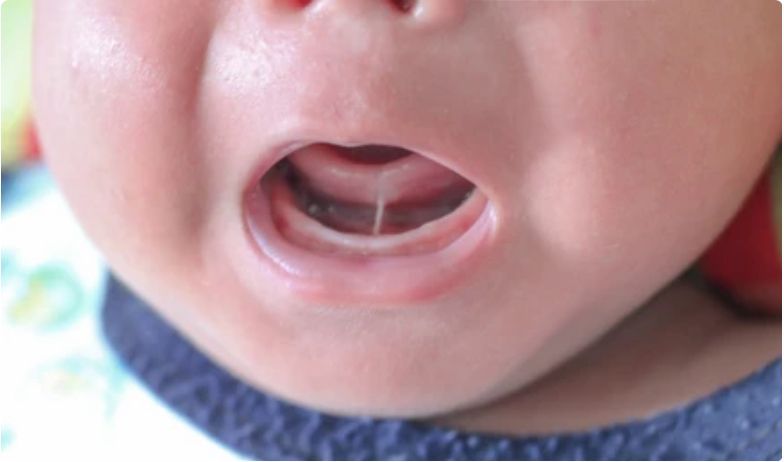
Understanding Tongue Tie in Newborns: Expert Recommendations and Treatment Options for a Healthy Breastfeeding Journey

Child passenger safety has evolved and improved dramatically in the past decade. Current estimates indicate that child safety seats and boosters reduce the risk of injury by up to 80% compared to children in seatbelts. Despite this progress, thousands of young children in our country are injured or killed in motor vehicle crashes each year. The American Academy of Pediatrics offers these best practices to optimize your child’s safety while traveling in a traditional motor vehicle: INFANTS AND TODDLERS should ride in the back seat, in a rear-facing infant or convertible car seat as long as possible, until they reach the highest weight and height allowed by the car seat manufacturer. Optimally this is age 2 years, weight up to 35lbs, or height up to 35 inches, depending on the model. Additional safety features available in some rear-facing car seats include load legs and anti-rebound bars to absorb the energy of a crash and reduce rotation. (Some convertible seats have higher weight limits.) CHILDREN who have outgrown their rear-facing car seat should continue to ride in the back seat, and now use a forward-facing convertible or combination car seat with a harness for as long as possible, up to the highest weight and height recommended by the manufacturer. Optimally this is age 4 years, weight at least 40lbs or height at least 40”. CHILDREN who have outgrown their forward-facing car seat should continue to ride in the back seat, and now use a belt-positioning booster seat until the vehicle lap and shoulder seat belt fits properly, typically when they have reached 4’9” (57”) and age between 8-12 years. When CHILDREN are old enough and large enough to use the vehicle lap and shoulder seat belt alone, they should always be belted in for optimal safety. Remember: the safest place to ride for ALL CHILDREN YOUNGER THAN 13yr is the back seat. CHILDREN WITH SPECIAL NEEDS (prematurity, altered muscle tone, decreased neurological control, skeletal abnormalities, airway compromise, etc.) may require specialized child restraint systems for optimal safety. Products are listed on the website for the National Center for the Safe Transportation of Children with Special Health Care Needs. When shopping for a car seat, keep in mind: No one seat is the “best” or “safest.” All car seats for sale in the USA have been crash-tested and must meet federally-approved standards. The best seat is the one that fits your child’s size, is correctly installed, fits well in your vehicle, and is used properly every time you drive. Do not decide by price alone. A more expensive car seat does not mean the seat is safer or easier to use. Avoid using a seat if you do not know its history (seats that may have been damaged in an accident are no longer safe). Never use a car seat that is old, cracked, does not have a label with the date of manufacture and model number, does not have instructions, or has been recalled (you can find out by contacting the National Highway Traffic Safety Administration Vehicle Safety Hotline at 888-327-4236 or check the website www.safercar.gov ). Car seats may be installed using either the vehicle’s seat belt or its LATCH system (lower anchors and tethers for children). Even if you have experience with installing and using car seats, it is important to read the vehicle owner’s manual and the car safety seat manual each time you install a new seat. Many communities have child passenger safety technicians who have completed a standardized National Highway Traffic Safety Administration course, and who can provide hands-on advice and guidance to families to ensure the child seat is properly installed. Resources available in our community include: Coral Springs Fire Department . 2801 Coral Springs Dr. Coral Springs, FL 33065 Phone: 954.344.5934 contact: Fire Administration ( Coral Springs and Parkland ) Broward Sheriff's Office/Tamarac. 7515 N Pine Island Rd. Tamarac, FL 33321 Phone: 954.720.2225 Tamarac Fire Department . 6000 Hiatus Rd. Tamarac, FL 33321 Phone: 954.597.3800 Boca Raton Fire Rescue Services Department . 6500 Congress Ave Boca Raton, FL 33487 Phone: 561.982.4000 Plantation Fire Department . 8101 W Broward Blvd. Plantation, FL 33324 Phone: 954.797.2150 Broward Sheriff's Office Weston . 17350 Royal Palm Blvd. Weston, FL 33326 Phone: 954.389.2015 Pembroke Pines Police Dept . 9500 Pines Blvd Pembroke Pines, FL 33024 Phone: 954-436-3274 Contact: Community Affairs Unit Prenes Chevelon Memorial Hospital Miramar . 1901 SW 172nd Ave Miramar, FL 33029 Phone: 954.538.5180

As a parent of an adolescent, you are in the best position to talk with your child regarding the importance of informed and healthy choices regarding their behaviors, including those around sex. The American Academy of Pediatrics (AAP) and your pediatricians recommend you discuss abstinence (not having sexual intercourse), as well as reliable contraception and condom use to prevent sexually transmitted infections (STIs) before your adolescent prepares to become sexually active. The data are clear: almost 25% of female high school students in the U.S. are sexually active. Of these, less than one-third are using an effective method of birth control, and less than one-half are using a condom. The risk of unintended pregnancy over the course of one year in couples who do not use any method of contraception is approximately 85%. Prevention of unintended pregnancy using effective contraception in sexually active adolescents is therefore paramount . Your willingness to discuss, sanction and provide access to contraception removes many of the barriers to your adolescent obtaining birth control. There are many factors for the adolescent and engaged parent to take into consideration when choosing the best contraceptive method. These include effectiveness, the reality of compliance, and potential side effects given the underlying health of the adolescent and family. For most adolescents, the benefit of any method of reversible contraception outweighs potential risk, though absolute contraindications to hormonal birth control do exist and will be discussed. Here is the landscape of available contraception. Long-acting reversible contraceptives (LARC) are the most effective reversible methods of contraception. The pregnancy rate is <1 percent per year in typical patients using this method, and fertility returns quickly after removal. LARC devices are inserted into the uterus (intrauterine devices, or IUDs), and once in place, do not require any action on the part of the adolescent. These are considered first-line options for adolescents by the AAP. LARC options include the non-hormonal copper IUD (e.g. PARAGARD), and the hormonal IUD (e.g. MIRENA) which releases the progestin hormone levonorgestrel. Contraceptive effects of the copper IUD last 10+ years, and contraceptive effects of the hormonal IUD last from 3-5 years. Noncontraceptive benefits of the hormone-releasing IUDs may include reduction in heavy menstrual bleeding, menstrual discomfort, as well as suppression of menses. Possible side effects of the copper IUD include cramping and spotting. Possible side effects of the hormonal IUD include headache, acne, breast soreness, mood changes, and irregular bleeding. The only contraindications of using IUDs for long-acting contraception include severe distortion of the uterine cavity, active pelvic infection, known or suspected pregnancy, Wilson disease (for the copper IUD), and unexplained vaginal bleeding. Contraceptive implant is another type of LARC. It is a single flexible plastic rod placed under the skin of the upper arm, which releases the hormone etonogestrel to prevent ovulation (e.g. NEXPLANON). Contraceptive effects last up 3-5 years. It is an attractive option for adolescents who desire long-term, uninterrupted birth control, and once placed, requires no action on the part of the adolescent. The implant is as effective as the IUD, with pregnancy rates < 1% per year. Possible side effects include spotting, weight gain, headache, nausea, and breast pain (side effects diminish over time). The contraindications of using the contraceptive implant include known or suspected pregnancy, abnormal vaginal bleeding, and lupus. Depot medroxyprogesterone acetate (DMPA)(DepoProvera) is an injectable hormonal (progestin-only) contraceptive that provides effective, reversible contraception for three months. Noncontraceptive benefits of DMPA include protection against ovarian cancer and endometrial cancer, ectopic pregnancy, benign breast disease, acne, and iron deficiency. The pregnancy rate is around 5% per year in typical patients. Possible side effects include weight gain, menstrual changes, headache, dizziness, acne, breast swelling, and changes in mood and libido. Adolescents using DMPA, whose reproductive plan includes pregnancy, ought to know that fertility may be delayed for more than a year following its use for contraception. Though unlikely in adolescents, contraindications for using DMPA include multiple risk factors for cardiovascular disease (smoking, diabetes, high blood pressure, high cholesterol) and lupus. Sides effects of DMPA may include unscheduled bleeding and decreased bone mineral density (reversible). Oral contraceptive pills (OCPs) have been FDA-approved for use for over 60 years. Currently, there are three different types of OCPs available on the market: the combination pill (estrogen and progestin, e.g. YAZ and LOESTRIN, used for 21 or 24 days depending on the brand), the progestin-only pill (norenthindrone e.g. MICRONOR, and norgestrel e.g. OPILL, used for 28 days), and the continuous use pill (estrogen and progestin, e.g. SEASONIQUE and LYBREL, used continually for 84 days or up to 365 days). The pregnancy rate with these methods is about 5% per year in typical patients. Oral contraceptive pills require the adolescent to take a pill daily and to remember to refill their prescription. The progestin-only pill needs to be taken at roughly the same time every day, has no hormone-free interval, and its main side effect is irregular bleeding and spotting. Noncontraceptive benefits of the progestin-only pill include reduced bleeding and less painful periods, and they are safe for people with high blood pressure, migraine, and those at risk for blood clots (see below). Noncontraceptive benefits of combined hormonal contraception include improved bone density and protection against ovarian cancer, endometrial cancer, ectopic pregnancy, benign breast disease, ovarian cysts, acne, PMS and premenstrual mood disorders, and iron deficiency. Possible side effects of the combined OCP include breakthrough bleeding, nausea, headaches, abdominal cramping, breast tenderness, increased vaginal discharge or decreased libido. Nausea can be avoided by taking the medication at night before sleep. Contraindications for using combined OCPs include multiple risk factors for cardiovascular disease (smoking, diabetes, high blood pressure, high cholesterol); migraine with aura; known mutations that predispose to blood clot formation; and lupus. FYI: OPILL is the only FDA-approved, daily oral contraceptive, progestin-only, now available over the counter (without a prescription), without age restriction, in stores and online. Transdermal contraception patch is a combined hormonal patch, containing both estrogen and progestin (ethinyl estradiol and norelgestromin, e.g. XULANE, or ethinyl estradiol and levonorgestrel, e.g. TWIRLA). It is applied weekly to the skin (at a different site) for three weeks, followed by a patch-free week (during which menstrual bleeding occurs). The contraception patch requires the adolescent to follow careful instructions in applying the patch, remember to change it weekly, and refill their prescription in a timely fashion. The pregnancy rate is around 5% per year in typical patients. Nonhormonal side-effects of the contraceptive patch include application site reactions (itching, irritation, redness) and true allergic reactions. In addition to the general contraindications for estrogen-progestin contraception described above, obesity (body mass index BMI >30 kg/m- or ≥95th percentile for age) is a contraindication for transdermal contraceptive patches: there appears to be decreasing patch effectiveness with increasing BMI, and there may be an increased risk of blood clots. Vaginal contraception ring is a vaginal ring (e.g. NUVARING) that has combined progestin and estrogen hormones (etonogestrel/ethinyl estradiol) that prevents ovulation. It is inserted into the vagina, left in place for three weeks, and removed for a single ring-free week (during which time menses occurs). The pregnancy rate is around 5% per year in typical patients. Adolescents who choose the vaginal ring must be comfortable self-inserting it into the vagina. Noncontraceptive benefits include lighter periods, improved cramps, and reduced acne. Side effects may include headaches, nausea, vaginal discharge, breast tenderness. Contraindications are similar to the combination OCP. Condoms are made of latex or synthetic material, designed to fit over the penis, and prevent introduction of sperm and ejaculation fluid into the vagina. Condoms are effective for pregnancy prevention and protection against STIs, as long as they are used properly. (NOTE: Natural fiber condoms will not provide protection against STIs.) With consistent and correct use of condoms, the pregnancy rate is around 2% per year, though with typical use, it is estimated to be about 15% per year (increased rate due to slipping off, breaking, etc). Other barrier methods include diaphragms, cervical caps, and sponges, though they are less effective (pregnancy rates 15-20% per year), they require conscious action of the part of the adolescent and/or partner at the time of sexual intercourse, and they do not protect against STIs. Contraceptive gel and spermicides are gels, creams, or foams applied inside the vagina before having sex. They contain chemicals that are toxic to sperm. They have no hormonal side effects, though they are less effective in preventing pregnancy than all the other methods discussed (pregnancy rates 20% per year), and they do not protect against STIs. Emergency contraception (EC) refers to an immediate intervention that prevents pregnancy from occurring after an episode of unprotected intercourse. Unprotected intercourse can be a result of contraception misuse, nonuse, or can result from forced sexual activity. EC is recommended within 5 days of unprotected sex. The available types of emergency contraception are the hormone ulipristil acetate 30mg x1 dose (e.g. ELLA), the hormone levonorgestrel 1.5mg x1 dose (e.g. PLAN B ONE STEP, available over the counter without a prescription), combined OCPx 2 doses over 12 hours, and emergency insertion of the copper IUD. Side effects from the use of EC pills are similar to those of oral contraceptive pills, such as nausea and vomiting, slight irregular vaginal bleeding, and fatigue. In addition to the use of contraceptives for birth control, there are medical conditions for which OCPs may be indicated . These include heavy menstrual bleeding, painful periods, iron deficiency anemia, irregular menstrual bleeding, acne, and polycystic ovarian syndrome (PCOS). Special circumstances in which contraception may be indicated include adolescents with physical or intellectual disability, for whom menstrual hygiene and menstrual discomfort may be challenging. For such adolescents, hormonal contraception (e.g. levonorgestrel -releasing IUD, DMPA, the contraceptive patch, or continuous or extended cycles of combined oral contraceptive pills) may be beneficial. We are here for you. Reach out on the portal or schedule an appointment with your adolescent to come see us in the office to discuss contraception options . Here are some additional resources and community partners that are available to you: Memorial Healthcare Adolescent Gynecology Dr. Elba Iglesias 954-265-1460 Omega Women’s Care Gynecologist Dr. Brooke Slaton 954-755-1411 Gynecologist Dr. Lona Sasser 954-340-1050 Contraception Explained: Birth Control Options For Teens & Adolescents https://www.healthychildren.org/English/ages-stages/teen/dating-sex/Pages/Birth-Control-for-Sexually-Active-Teens.aspx Healthy Teens | ACOG Bedsider Birth Control Support Network Center for Young Women's Health Planned Parenthood | Official Site

Caring For Your Child With Obesity

Postpartum mood disorders are common medical conditions that can significantly impact the heath, well-being, and connection of mothers, infants, and their families. They are also a leading cause of maternal mortality in the USA. The acronym used to encompass postpartum mood disorders is PMADs, short for postpartum mood and anxiety disorders, though they include a whole spectrum of postpartum mood disorders. These include postpartum depression (low mood, sadness, feelings of helplessness and hopelessness); postpartum anxiety (constant worrying, intrusive thoughts, inability to turn off the constant chatter in the mind); postpartum obsessive compulsive disorder (OCD)(obsessive/unwanted thoughts and fears, ritualistic behavior, avoidance of triggering stimuli); postpartum post-traumatic stress disorder (PTSD)(tension, sleep disorder, flashbacks of a traumatic event regarding pregnancy/pregnancy loss/childbirth/neonatal death); and postpartum psychosis (delusions, hallucinations, paranoia, suicidal or homicidal thoughts). PMADs are estimated to affect up to 25% of all moms and birthing parents, making it the most common under-diagnosed complication of pregnancy. In the height of the COVID-19 pandemic, up to 40% of moms and birthing parents met the criteria for a postpartum mood disorder. PMADs disproportionately affect moms of color and those living in poverty (up to 50%). LGBTQIA+ moms and birthing parents are also at a higher risk for developing a PMAD. PMADs can occur with the onset of delivery or anytime within the first four months of delivery, and last a year or longer. Risk factors for the development of PMADs are a personal history of anxiety or depression, young maternal age, social isolation and lack of support, financial stress, previous pregnancy complication or loss, traumatic birth, partner-related stress, substance abuse, negative early breastfeeding experiences, and family history of postpartum mood disorder. It is normal for most new moms to transiently experience “postpartum blues,” a brief period after delivery characterized by emotional lability, exhaustion, weight loss, anxiety, intrusive thoughts, trouble sleeping, loss of interest in usual activities, and low mood. PMADs must be considered and addressed when these symptoms occur daily, persist longer than a few weeks after delivery, and cause significant impairment in function. PMADs can impact bonding between mother/birthing parent and infant. PMADs can interfere with breastfeeding and all forms of nourishing the infant. Lack of maternal interest, attention and attachment due to PMADs can result in safety issues for the infant, as well as emotional deprivation, poor behavioral regulation, failure to thrive, sleep disorders, developmental delay, increased risk of abuse and neglect, and subsequent mood disorders in their later childhood, adolescent and adult lives. Paternal PMADs are also of note, affecting up to 15% of dads in the first 6 months following delivery. Paternal PMADs may present differently, with substance abuse, domestic violence, and compulsive behaviors. Paternal PMADs can significantly impact relationships with their partners, and increase the risk of developmental, behavioral and psychiatric disorders in their children. It is so important that postpartum mood disorders be identified and addressed in all affected parents. Screening for PMADs can be instituted during visits to the pediatrician and the OB-GYN. Since the first OB-GYN visit is generally not until 6-8 weeks postpartum, pediatricians are in a unique situation to screen for maternal PMADs at the 1 month well-child check. The 10-question Edinburgh Postnatal Depression Scale (EPDS) is the validated screening tool most commonly used, and available in many languages. A score of 10 or higher raises the concern of a PMAD and ought to trigger referral to resources for support, diagnosis and treatment. A score of 20 or higher raises the concern of severe depression or psychosis and ought to trigger immediate referral for an emergency psychiatric evaluation. As already discussed, there are significant consequences to mom, infant, and their family if PMADs are left untreated. Despite the consequences of untreated PMADs and the presence of a range of options for effective therapy and treatment, most mothers/birthing parents with postpartum depression and anxiety do not seek treatment. There are many factors at play, including moms not recognizing what they are feeling is common and normal; the stigma of mental illness, particularly maternal mental illness; social isolation with COVID; the societal expectation that being a new mom is a happy, joyful experience, and the guilt/shame many moms feel when they are having a different experience; the fear that admitting thoughts/feelings of postpartum depression or anxiety could lead to the baby being taken away from the mom. Screening for perinatal and postpartum depression normalizes these conditions, illuminates their prevalence and their importance. Yet the great majority of moms suffering from PMADs fail to be screened and diagnosed, and are thus left untreated. Treatment for PMADs includes home visits, emotional support groups, exercise, relaxation techniques, psychotherapy, natural treatments like fish oil, and pharmacological therapy when indicated. In moderate to severe postpartum depression and anxiety, the benefit of using medication far outweighs any risk to the baby while breastfeeding. Treating PMADs with appropriate medication is generally safe for the baby and does not preclude breastfeeding . Medications most often used to treat postpartum depression and anxiety are SSRIs, which block serotonin reuptake, increasing the level and availability of this “happy hormone” at the nerve synapse, improving the symptoms of depression, anxiety and OCD. Sertraline (Zoloft) is the best SSRI for breastfeeding moms, as it has the lowest concentration in breast milk and thus the lowest risk for side effects in the infant (restlessness, irritability, colic, poor feeding). SNRIs like duloxetine (Cymbalta) block the reuptake of norepinephrine at the nerve synapse: they are another option in the safe, effective treatment of PMADs. When treatment of the mother requires medication that is unsafe while breastfeeding, other feeding options should be explored, as the health and well-being of the mother must be the first priority. Postpartum psychosis is a rare but serious PMAD, occurring in approximately 1/1000 deliveries. Moms and birthing parents with postpartum psychosis may demonstrate wide mood swings, irritability, paranoia, hallucinations, delusions, and suicidal or homicidal thoughts, placing them and their babies at risk for harm. Risk factors include personal and family history of bipolar depression and schizoaffective disorder. Hormonal shifts, sleep deprivation, environmental stress, and stopping mod-stabilizing medications are believed to be contributing factors. Postpartum psychosis is a medical emergency, often requiring hospitalization for stabilization and treatment. We are here for you. If you have concerns about postpartum depression or anxiety and its impact on your ability to care for yourself and your child, reach out on the portal or schedule an appointment to come see us in the office. Here are some additional resources and community partners that serve our moms and birthing parents with postpartum mood disorders: Psychiatry/Women’s Health Dr. Nicole Derish. www.derishmd.com . 786-471-6132 Therapy/Women’s Health Dr. Laura Meyer www.expeditionsinmotherhood.com 954-884-0050 Therapy/Maternal Mental Health Dr. KC Charette 561-617-3323 Therapy/Women’s Health Karyn Rosenberg, LCSW, PMH-C 561-306-0232 Therapy/Maternal Mental Health Sarah Hendin, LMHC 561-542-4709 Therapy/Women’s Health Laura Kreisler, LCSW 561-376-0164 Broward Health Coral Springs Postpartum Support Group Contact Lisa Hayes 954-346-4260 or Barbara Bolinsky 954-344-2229 Holding Space for New Mamas Support Group 786-829-5950 Broward Healthy Start Coalition. www.browardhsc.org . Call Connect 954-567-7174 Healthy Mothers, Healthy Babies Coalition: Mothers Overcoming Maternal Stress (M.O.M.S.) www.hmhbbroward.org . 954-765-0550 ext.331 www.themotherhoodcenter.com 212-335-0034 Postpartum Support International PSI. www.postpartum.net 1-800-944-4773 or text “Help” to 800-944-4773 SAMHSA 24/7 National Hotline: 800-662-HELP (4357) www.2020mom.org . 310-955-1080 www.healthynewmoms.org 570-955-7821 or text HealthyMOMS to555888 www.postpartummen.com . 415-346-6719 DrWill@TheMensDoc.com Early Steps www.Floridaearlysteps.com 954-712-0200 Henderson Behavioral Health www.hendersonbh.org 954-606-0911 24/7 Crisis 954-463-0911

Iron is a mineral required for numerous critical functions in the human body, including oxygen transport, gene regulation, DNA synthesis, brain function, and musculoskeletal function. Iron is ingested in various forms from food it our diet, and absorbed from the small intestine. Iron is stored as heme in red blood cells (hemoglobin) and muscle cells (myoglobin); iron is stored as ferritin in the liver, spleen, skeletal muscle and bone marrow; and to a smaller extent, iron is stored as hemosiderin deposited in various tissues. Iron deficiency refers to a state in which there is insufficient total body iron to maintain normal physiologic functions. Though the body efficiently recycles iron from aging blood cells, iron deficiency may result from increased iron need, particularly during rapid periods of growth, in the face of deficits in the ingestion or absorption of iron, or from excessive iron utilization or iron/blood loss. The impact of Iron deficiency is monumental. Iron deficiency, independent of anemia, is estimated to affect as many as 50% of young children and female adolescents worldwide. In the United States, approximately 15% of toddlers and 5% of preschool children are iron deficient. In America, the prevalence of iron deficiency is higher among children living at or below the poverty line, and compared to white children, the prevalence of iron deficiency is higher in young Hispanic/Latin American children, in Asian American children, and children from families recently immigrating to the United States. This is of huge consequence, as iron deficiency is associated with lower IQ scores, lower assessments of emotional health, attention deficit hyperactivity disorder (ADHD), restless leg syndrome (RLS), visual and auditory deficits, immune dysfunction, impaired muscle metabolism, chronic fatigue, breath-holding spells, and thyroid dysfunction. These impairments in neurodevelopmental, cognitive, immune, and musculoskeletal function may occur prior to the diagnosis and treatment of iron deficiency anemia. Iron deficiency is the most common nutritional deficiency in the world and results from a wide variety of causes. Inadequate dietary intake of iron-rich foods, the consumption of foods that impair iron absorption, and menstruation are the main contributing factors. Iron is stored prenatally, and iron stores are generally adequate in full-term infants until age 3-4 months. Preterm infants, infants with intrauterine growth retardation, infants of diabetic mothers, infants of moms with iron deficiency, infants who experience any hemorrhage/blood loss, and breastfed infants are at higher risk for anemia at 3 months, and should be started on iron supplementation if screening is positive for anemia. By 6 month age, infants are able to ingest iron-rich and iron-fortified foods, including fortified cereal, leafy greens like spinach, beans and lentils, liver, red meat, etc. Iron deficiency may develop in young children and adolescents who are pickey eaters; those who avoid animal products; those who consume excess (>24 oz per day) unfortified cow milk or goat milk; and those who ingest excess tannates in tea, phosphates in brain rich foods, and phytates in plant fiber, especially in seeds and grains. Those on a vegan diet are particularly at risk for iron deficiency. In addition, iron deficiency may result from excess blood loss from heavy menstruation (indicators include soaking of a pad in less than 2 hours, bleeding into clothes, or blood clots larger than 1 inch); and excess loss of blood though the gastrointestinal tract (e.g. colitis from milk allergy, inflammatory bowel disease, polyps). Iron deficiency can result from inadequate absorption of iron due to diseases of the GI tract, including celiac disease, inflammatory bowel disease, H. Pylori infection, giardiasis, and autoimmune gastritis. Overweight and obese adolescents are at risk for iron deficiency: compared to the 3% incidence of iron deficiency for adolescents with a normal BMI, overweight adolescents with a BMI of 85%-95% have an incidence of iron deficiency of 7%, and obese adolescents with a BMI over 95% have an incidence of iron deficiency of close to 10%. By complex mechanisms, athletic adolescents both utilize iron and lose iron at a higher rate, placing them at risk for iron deficiency. Finally, there are genetic mutations that result in the failure of iron absorption, even in the presence of iron deficiency, causing persistent iron deficiency resistant to oral iron supplementation. Clinical manifestations of iron deficiency are generally mild: in fact, most infants and young children with iron deficiency are asymptomatic and found on routine labs to have a microcytic (small cell) anemia. Much less frequent are infants and children with severe anemia, who present with lethargy, pale skin, irritability, poor feeding, rapid heart rate and rapid respiratory rate, and exercise intolerance. Children with iron deficiency may exhibit pica, the intense craving for nonfood items, such as clay or dirt, rocks, starch, chalk, soap, paper, and cardboard. Craving for ice is particularly common and specific for children and adolescents with iron deficiency anemia. Evaluation for iron insufficiency and iron deficiency anemia includes a complete blood count (CBC), measures of serum iron and iron binding capacity, and iron storage (ferritin). Iron deficiency is often defined as a serum ferritin < 15 mcg/L in all pediatric age groups, noting that ferritin will be decreased with iron depletion prior to the onset and diagnosis of iron deficiency anemia. As the production of blood cells is restricted by iron insufficiency, the hemoglobin will start to decrease, and the volume of the red blood cells (MCV) will diminish, though true anemia may not yet have occurred. Iron deficiency anemia (IDA) in children is defined in children 6 months to <5 years as ferritin <15 mcg/L and hemoglobin <10.5 g/dL; in children 5 to <12 years ferritin <15 mcg/L and hemoglobin <11.5 g/dL; and in adolescents older than 12 years ferritin <15 mcg/L and hemoglobin <12 g/dl. Differential diagnosis includes thalassemia trait, a genetic variation in hemoglobin that causes the blood cells to mature rapidly and leave the bone marrow sooner, at a relatively smaller size (low MCV). Lead toxicity can also present with microcytic anemia. Anemia of chronic disease will also cause blood cells to be smaller, and serum iron to be lower, though inflammatory markers and ferritin are generally elevated. At Children’s Medical Association, we check a CBC to screen for iron deficiency at 3 months, 12 months, and yearly at well child check-ups. We screen for lead toxicity between 12-24 months. Additional screening is targeted for those with heavy menstruation, those on a vegan diet, those with chronic disease, and others at high risk. Management and treatment of iron deficiency starts with dietary interventions to improve iron intake. Increase foods rich in iron: animal products such as red meats, liver and pork, legumes such as beans and lentils, dried fruits (prunes, raisin, apricots), leafy greens (spinach, kale, Swiss chard), and iron-fortified cereals. Foods rich in vitamin C (e.g. citrus fruits, cantaloupe, strawberries, tomatoes, and dark green vegetables) are recommended to enhance iron absorption. Reduce consumption of beverages such as tea and soda that can affect iron absorption. Limit cow milk consumption to 24oz per day. For infants and children with a presumptive diagnosis of iron deficiency anemia based on the history and initial laboratory testing, the next step is a therapeutic trial of iron, consisting of ferrous sulfate, 3 mg/kg of elemental iron, once daily in the morning or in divided doses between meals. NovaFerrum is a recommended iron supplement which is allergen-free, tastes great, and is available in 15mg/ml and 25mg/ml of iron per dose. Other products include Vitamin Friends Iron Gummies (15mg), Natural Factors Easy Iron Chewable (20mg) and Prothera Iron Chewables (30mg). Iron may be given alone or with water, juice, or acidic fruits (e.g., mango, strawberries, or applesauce). Milk and/or dairy products should be avoided for approximately one hour before and two hours after each dose, as the calcium interferes with iron absorption. For adolescents with iron deficiency, with or without anemia, we recommend ferrous sulfate, providing 65 to 130 mg elemental iron daily. Typically, patients treated for iron deficiency anemia return to the office 2-3 months later for a repeat CBC, monitoring the change in hemoglobin and MCV. Treatment courses should last for at least 3 months, and even after discontinuation of iron therapy, it is reasonable to continue iron-rich foods in the diet. Transfusion therapy is rarely needed for iron deficiency anemia in children and adolescents, unless the hemoglobin concentration is below 7 g/dl. Indications for IV iron therapy include persistent anemia with oral iron intolerance, malabsorption, or nonadherence to oral iron therapy. Transfusions are generally reserved for patients who are in distress due to anemia, with rapid heart rate, rapid respiratory rate, low blood pressure, light-headedness, and lethargy. Children with underlying gastrointestinal disease, such as short bowel syndrome or inflammatory bowel disease, may have particular difficulty tolerating oral iron and require early initiation of IV iron therapy. Prevention of iron deficiency is supported by a well-balanced diet, including iron-rich foods detailed above. For those at increased risk for iron deficiency, more frequent screening and iron supplementation are both tools to prevent deficiency. There has been much attention to the idea of delayed cord clamping as a means of providing more placental blood and iron to the neonate, as a means to prevent iron deficiency later in infancy. Research has shown that delayed cord clamping of greater than 2 minutes leads to elevated ferritin levels and decreased risk of iron deficiency in the infant up to 8 months of age compared with early cord clamping (5-10 seconds). We are here for you. If you have concerns about your child and iron deficiency, reach out on the portal or schedule an appointment to come see us in the office. "This message is intended only for use by the person or entity to which it is addressed. Because it may contain confidential information intended solely for the addressee, you are notified that any disclosing, copying, downloading, distributing or retaining of this message, and any attached files, is prohibited and may be in violation of state or federal law. If you received this message in error, please notify the sender by reply mail and delete the message and all attached files."
Infantile hemangiomas are “birth marks” made up of a collection of blood vessels that formed incorrectly and proliferate rapidly. They occur in as many as 5% of infants, making them the most common benign tumor of infancy. Infantile hemangiomas are more common in girls, twins, white infants, and those born premature or with low birth weight. Most are small, resolve on their own, and require no treatment. A small number of hemangiomas have the potential to cause problems, based on their size or their location, and those risk factors are important in the consideration for treatment. Careful monitoring, recognition of risk, and early intervention are key in providing the best care for your infant with hemangioma. Infantile hemangioma may be present at birth as a superficial, small, red, vascular mark on the skin, or more commonly become evident in the weeks following birth. They have traditionally been called “strawberry hemangiomas.” There is a natural history of uncomplicated infantile hemangiomas : they typically grow rapidly during the first 3 months, most stop growing by 5 months, and most infantile hemangiomas start to involute between 5-12 months of life. The color will change from red to milky-white or gray, the lesion will flatten and shrink, and 90% heal by age 4yr. Even after involution, the skin may be left with thin blood vessels (telangiectasias), redundant skin, or a scar. Some infantile hemangiomas are deeper in the skin, and may have a blue appearance. As stated above, most infantile hemangiomas are benign. There are well-defined categories of infantile hemangiomas with increased risk for complication : their early recognition and treatment can prevent adverse outcomes. Hemangiomas requiring treatment include: Those in the “beard-area” or the front of the neck, which may be associated with hemangioma in the airway, which has the potential to block the airway as the hemangioma grows. Infants with 5 or more cutaneous hemangiomas, which may alert to the possibility of hemangiomas in vital organs like the liver, and may cause strain on the body leading to cardiac failure and severe thyroid dysfunction. Infantile hemangiomas in areas where their presence may cause functional impairment, e.g. those at the eye potentially impacting vision, and those at the lip/mouth potentially impacting feeding. Infantile hemangiomas in areas at increased risk for ulceration/bleeding, e.g. those at the lip, upper ear, diaper area, anus, and skin folds of the neck, arm pit, and groin. Infantile hemangiomas in areas at high risk of scarring or permanent disfigurement, e.g. those at the face and scalp, those at the breast, those with a steep step off (ledge effect), and those greater than 2cm in size or 2mm in depth. Large, segmental (greater than 5cm) infantile hemangiomas over the head and neck, or lumbosacral area of the back, which may be associated with underlying structural anomalies of the brain, vascular system, spine or spinal cord. It is recommended that infants with hemangiomas that are high risk for complications be seen by a specialist by 1 month of age. Depending on the location and nature of the hemangioma, specialty consultation may be arranged with a pediatric dermatologist, a pediatric plastic surgeon, and/or an otolaryngologist (ENT surgeon). Diagnostic studies are generally unnecessary, as infantile hemangiomas are diagnosed by their clinical appearance. Rarely, if the appearance or growth or response to treatment are atypical, a biopsy may be recommended. Ultrasound of the abdomen is indicated in the setting of 5 or more cutaneous hemangiomas, to evaluate for the presence of hemangiomas within the liver. The presence of large segmental hemangiomas, for example over the head and neck or lumbosacral spine, would warrant imaging studies (MRI/MRA) of associated structures like the head, chest, spine and spinal cord. The first-line treatment of high-risk infantile hemangiomas is oral propranolol. Studies show a mean response of 95%. The precise mechanism of action is unclear, though may be related to its ability to promote vasoconstriction and inhibit angiogenesis (new blood vessel formation). The recommended starting dose is 0.6 mg/kg given twice daily, with a gradual increase to the target dose of 2-3 mg/kg/day. Treatment is recommended for 6 months, and there may be rebound growth in 10-25% of infants when propranolol is discontinued. Propranolol is a beta-blocker, and side effects of propranolol include bradycardia (slow heart rate), hypotension (low blood pressure), hypoglycemia (low blood sugar), lethargy, poor feeding, wheezing, and sleep disturbance. Any child with known congenital heart disease and hemangioma should be seen by their cardiologist for consultation, regarding the safety of propranolol with their underlying cardiac condition. Screening EKG is only indicated for infants with a baseline low or irregular heart rate, and those with a family history of congenital heart disease or rhythm disturbance. Propranolol should be administered with or after feeds, and should be held at times of diminished feeding or vomiting to reduce the risk of hypoglycemia. Side effects , when they occur, are generally mild, and may be managed by lowering the dose of propranolol. Discontinuation of treatment is rarely necessary. For patients in whom propranolol is contraindicated, poorly tolerated or ineffective, second-line treatment of high-risk infantile hemangiomas is steroids, either orally or by direct injection into the high-risk lesion. Prolonged use of oral steroids is associated with significant side effects (Cushingoid appearance, increased risk for infection, poor growth, high blood pressure, and mood changes), and is thus not recommended. Low risk lesions do not require treatment , though some clinicians will recommend treating thin, superficial hemangiomas with topical timolol, also a beta-blocker, to slow growth and enhance resolution. Lesions treated with topical timolol have an estimated clearance of over 60%. Since medication may be absorbed through the skin into the bloodstream, a small percentage of infants treated with topical timolol may experience systemic symptoms, including sleep disturbance, bradycardia, and wheezing: these side effects are noted to be more prevalent in premature infants. Surgical interventions during infancy are generally avoided (risk of anesthesia and bleeding), though high-risk lesions that are not responsive to oral propranolol may require surgery. Pulse-dye-laser (PDL) treatment is being used for infantile hemangioma, though it carries the risk of bleeding, ulceration, skin atrophy, and scarring. Both surgery and PDL may be useful in managing persistent skin changes that may result from an infantile hemangioma. We are here for you. If you have concerns about your infant’s hemangioma, particularly if it is rapidly growing between the 1 month and 2 month check-up appointments, reach out on the portal or schedule an appointment to come see us in the office. Here are some additional resources and community partners that serve our patients with infantile hemangioma: Plastic Surgeon Dr. Eric Stelnicki (Hollywood) 954-984-1899 Plastic Surgeon Dr. Drew Schnitt (Delray Beach) 855-467-7473 Plastic Surgeon Dr. Chad Perlyn (Miami) 305-278-5951 Dermatologist Dr. Lawrence Schachner (Miami) 305-243-6704 Dermatologist Dr. Ana Duarte (Miami) 305-669-6555 Dermatologist Dr. Anna Falabella (Hollywood) 954-961-1200 ENT Dr. Samuel Ostrower (Coral Springs) 954-265-1616 ENT Dr. Steven Singer (Hollywood) 954-987-5430 ENT Dr. Sandeep Dave (Miami) 786-624-3687 https://hemangiomaeducation.org/ Society for Pediatric Dermatology https://peds derm.net/

Concussion is a commonly occurring traumatic brain injury, which largely occurs in children and adolescents during sports-related trauma, though concussion may be related to other forms of head trauma (accidental falls, being struck by a person or object, motor vehicle injury etc). Up to 2 million recreational concussions and sports-related concussions are reported yearly in people under 18yr in the United States. Symptoms following concussion can impact physical, emotional, academic, social, family, and athletic health, making the appropriate management of your child with concussion extremely important, both enhancing recovery and minimizing long-term complications. Concussion may be caused by a direct blow to the head, or trauma elsewhere to the body that produces an energy force transmitted to the head, impacting the brain. The kinetics of the injury consist of forces of acceleration, deceleration, and rotation of the head. The trauma causes a chemical cascade and metabolic disturbance that impacts the brain down to the cellular level, depleting cellular energy, creating acidosis, causing oxidative stress and inflammation, all resulting in significant changes in brain function. The actual structure of the brain may not appear altered on standard Xray, CT, MRI. Decreased cerebral brain flow and the sheer force to the axons during trauma also impact brain function for days to weeks following concussive head injury. Signs and symptoms of concussion can be divided by category, and include: somatic (headache in over 90%, dizziness in up to 75%, nausea and vomiting, neck pain, sensitivity to light and noise) vestibular/oculomotor (visual tracking and convergence problems, impaired balance and coordination, slow reaction time, dizziness, hearing problems and tinnitus) cognitive (blank stare or “stunned” appearance, poor focus in over 50%, confusion and disorientation in 40%, slow processing speed, slow speech, brain fog, memory loss, impaired learning) emotional (irritability, emotional lability, sadness) sleep (drowsiness, fatigue, trouble falling asleep or staying asleep, sleeping too much) The sports with the highest risk of concussion are tackle football, lacrosse, ice hockey, wrestling, soccer, field hockey, and basketball. Rates of concussion are significantly higher during competition than practice. Interestingly, girls’ soccer and basketball are associated with a higher incidence of concussion than boys’ soccer and basketball. Swimming and track are the sports with the lowest rates of concussion. Concussion may also occur with accidental trauma during recreational activities such as bicycle riding, skateboarding, ice skating and skiing. Brief loss of consciousness in a child sustaining concussion occurs less than 10% of the time, and does not predict more significant impairment; the absence of loss of consciousness does not justify more rapid return to play or school. The initial evaluation of a conscious athlete who is suspected of having a sports-related concussion occurs on the field or on the sideline or in the locker room. The most common tool used is the Sport Concussion Assessment Tool (SCAT). Because athletes do not always show immediate symptoms or signs of deficits following trauma, it is imperative to err on the side of caution and keep the athlete from returning to play on that same day, while reassessing for evidence of concussion. In fact, all 50 states have enacted laws requiring the benching of an individual suspected of sustaining a concussion, and requiring medical evaluation before returning to play. Studies show that athletes who continue to play are nearly 10x more likely to have a prolonged recovery from this trauma, and may experience severe consequences following subsequent head trauma. Red flags in the initial assessment that would warrant urgent referral to an emergency room include loss of consciousness, weakness or tingling in the arms or legs, severe or progressive headache, repeated episodes of vomiting, irritability or combativeness, and/or seizure. These may indicate more serious trauma, such as skull fracture, hemorrhage, or injury to the cervical spine. CT scan of the brain is routinely performed in the emergency room for a child being evaluated for head trauma, though it is generally normal for the child with concussive head injury, in the absence of signs of more serious trauma. CT does expose a child to the potentially harmful effects of ionizing radiation, and ought to be considered carefully in a child who appears well and in whom there is no evidence for deterioration over time. MRI of the brain is superior to CT scan in detecting contusion (bruising) to the brain, and ruling out other brain lesions in children with ongoing symptoms (e.g. tumor or Chiari malformation). Functional neuroimaging (PET scans, functional MRI) may soon be available in the diagnosis and management of pediatric concussive head injury. Neurocognitive testing may be beneficial in the individual with prolonged symptoms or repeated concussion, though children and adolescents with underlying diagnoses of psychiatric illness, ADHD, migraine and substance abuse may have abnormal neurocognitive testing independent of concussion. The diagnosis of concussion is based on a history of trauma that results in rapid acceleration, deceleration and/or rotation of the brain; onset of signs of symptoms soon after the injury; a positive assessment, e.g. SCAT; exclusion of structural injuries to the brain, e.g. subdural hematoma noted on CT scan; the exclusion of other medical diagnoses with similar features, e.g., heat stroke, hypoglycemia, cardiac syncope, migraine, psychiatric disorder, etc. The acute management of concussion involves assessment for injuries/deficits that may require rehabilitation; education about what to anticipate following the diagnosis of concussion; cognitive and physical rest; and guidance for returning back to school and play. Current recommendations include: Immediate removal from play. Limiting initial physical exertion to walking, keeping the intensity below a level that provokes symptoms. Avoiding recreational activities that may result in a second head injury (cycling, skating, skiing, skateboarding, etc.) until full recovery. Suspending screen time for at least 24 hours following the injury, then limiting screen time in individuals with light sensitivity or oculomotor deficits. Consider accommodations such as enlarged font and lower brightness. Reducing academic workload to avoid provoking symptoms. Suspending driving for a few days following head trauma, as focus and reaction times may be impaired. Analgesics and anti-inflammatory medications like Tylenol and Motrin may be used in the days following injury, though prolonged use may result in rebound headaches and many complicate recovery. Ondansetron (Zofran) may be used for nausea in the first few days following concussion. However, ondansetron itself can potentially cause headache, drowsiness and dizziness, complicating other symptoms of concussion. Physical therapy may be useful to facilitate recovery when the concussion is accompanied by cervical strain or deficits in balance and coordination. Occupational therapy may be useful to facilitate recovery when the concussion is accompanied by deficits in motor planning and cognition. Oculomotor therapy may be useful to facilitate recovery when the concussion is accompanied by visual deficits in accommodation, tracking, etc. Recovery varies among all patients, as every concussion is unique, though most children and adolescents with concussive head trauma recover within 1-4 weeks. Return to sport must be individualized, and is best accomplished by following a graduated stepwise program. Studies have consistently showed that low levels of aerobic exercise, if tolerated, initiated within days of the injury, are safe, prevent deconditioning, and improve recovery time. Athletes should not be allowed to return to contact, collision or high-risk activities until symptoms of concussion have resolved and a return-to-sport progression has been completed. Each step should take at least 24 hours, so at best, it takes about 1 week to resume full participation, as long as the athlete remains symptom-free. The return-to-sport progression may be monitored by a parent, coach, athletic trainer, or health care provider working within a specialized concussion program. Written medical clearance from a health care provider is required for any athlete with concussion to return to participation. The process is rigorous, as athletes who return too soon are at risk for a more severe subsequent injury and prolonged recovery. Return to full participation is only recommended once your child is back to baseline, is symptom-free, and has successfully returned to school. Return to school must also be individualized, as symptoms of concussion may be worsened by cognitive effort (reading, prolonged screen time, high focus with problem solving and test taking, etc.). In addition, studies show that cognitive rest following concussion is associated with improved neurocognitive function. It is recommended to rest at least one to two days before returning to school, and then return to school when capable of sustaining 30-45 minutes of concentration without symptoms. For children and adolescents with ongoing symptoms, it is recommended to minimize cognitive activities; avoid exposure to video games, loud music and prolonged recreational screen time; and analogous to return to sport, gradually resume cognitive activities under the guidance of a concussion specialist. School adjustments may be necessary in the first few weeks (rest break in the nurses’ office, extra time in hallways, obtaining preprinted notes, etc.). Formal academic accommodations may be required to address longer-term needs beyond 3 weeks, and can be formalized in a 504 plan (extra time on work, changes in class schedule, alternate arrangements for standardized testing, etc.). Academic modifications may be necessary for prolonged post-concussive symptoms impacting school function, necessitating special education with needs specified in an individualized educational plan (IEP). The chronic management of concussion includes addressing long-term symptoms such as headache, dizziness, fatigue, brain fog, poor focus and memory, impaired balance and coordination, insomnia, poor emotional regulation, mood disorders, and sleep disorder. Referral to a specialized concussion center is warranted. Consultation with a pediatric neurologist, neuropsychologist, and/or physiatrist may be recommended. Physical therapy, occupational therapy, vestibular therapy, oculomotor therapy, biofeedback, and cognitive-behavioral therapy with a psychologist made be necessary. Good sleep hygiene is essential, and a trial of melatonin 2-5mg may be initiated to improve sleep. Though there are no evidence-based clinical studies to support nutritional strategies in the recovery from concussion, it makes sense to incorporate foods that reduce oxidative stress and inflammation, and support neuronal healing and growth: these include brightly colored fruits and vegetables, particularly blue and purple grapes and berries, and cruciferous vegetables like broccoli and cauliflower; wild salmon and bivalves (mussels, clams, oysters); healthy fats from eggs, nuts and seeds, avocado, coconut milk, olive oil; herbs like ginger, cilantro, and rosemary; spices like garlic, tumeric and cinnamon. Prevention of concussion is challenging , with efforts largely focused on improving protective equipment and their fit, implementing fair rules of the game to reduce risk of head injury, and educating participants and their families for safe sports participation. Policy change is also needed: based on clear evidence that concussion risk is reduced in youth ice hockey when body checking is prohibited, the American Academy of Pediatrics recommends that body checking not be permitted in youth ice hockey until age 15yr. Exercises done to strengthen neck musculature may mitigate the impact of an injury and reduce the risk of sports-related concussion. The prognosis for your child with concussion is good. Most pediatric patients will recover readily from concussion, and it is estimated that 90% will be symptom-free and clear to return to play within one month of the injury. The presence of a higher overall initial symptom burden has shown to be the most consistent predictor of prolonged (>28 days) recovery after a concussion. Additional risk factors for prolonged recovery include history of prior concussion, history of migraine, history of learning disabilities, history of anxiety or depression, and failure to observe the recommended periods of cognitive and physical rest. Female adolescent patients may take longer to recover. We are here for you. Reach out on the portal or schedule an appointment to come see us in the office if you have any concerns about your child with concussion. Here are some local resources to support our patients with concussion and their families: Dr. John W. Kuluz Nicklaus Children’s Hospital Concussion and Brain Injury Clinic 3100 SW 62nd Avenue Miami, FL 33315 786-268-1789 Drs. Michael Dressing, Diana Martinez, Virmarie Quinones-Pagan Joe DiMaggio Children’s Hospital Concussion Clinic 4651 Sheridan Street Suite 150 Hollywood, FL 33021 954- 538-5566 Sports Medicine Concussion Clinic Nova Southeastern University 3301 College Avenue Davie, FL 33314 954-262-5590 Head Strong Concussion Care Program Coral Springs Medical Center 3000 Coral Hills Drive Coral Springs, FL 33065 954-344-3180 University of Miami Concussion Program Batchelor Children’s Research Institute 1580 NW 10th Ave. 1st Floor Clinic Miami, FL 33136 305-243-2074 University of Miami Concussion Program UHealth of Boca Raton 3848 FAU Boulevard, Suite 305 Boca Raton, FL 33431 561-289-5808

Migraine is the most common acute and recurrent headache syndrome in children. Migraine prevalence estimates are around 3% of children by age seven years and about 10% by age 15 years. Before puberty, there is an equal prevalence in girls and boys, but after puberty, the prevalence is 2 to 3 times more common in girls. Early intervention is the key for managing migraine in children, to achieve fast and complete pain relief, to minimize the impact on physical and emotional health, and to promote rapid return to normal function, including participation in school, sports, family and social activities. The diagnosis of migraine is made with the occurrence of at least 5 headaches over the last year, lasting 2-72 hours if left untreated, with additional features including moderate to severe intensity, pulsatile quality, one sided, and worsening with activity. Migraine is often accompanied by aura (a sensation, whether visual or auditory or taste or smell, that precedes headache by 5-60 minutes), nausea, vomiting, abdominal pain, sensitivity to light (photophobia), sensitivity to sound (phonophobia), and generally relieved by sleep. Chronic migraine is defined as 15 or more headache days (8 migraines) per month for 3+ months. Triggers to migraine include environment (stress, weather changes, barometric pressure); lifestyle (stress, sleep disruption, dehydration, dietary triggers including food dye, MSG, aspartame, nitrates, caffeine, gluten); metabolism (mitochondrial dysfunction, related to energy production in our cells); hormones (neuroendocrine and menstrual, related to rapid changes in estrogen, serotonin, and inflammatory mediators like glutamate and prostaglandins). Identifying and avoiding these triggers are important steps in helping to care for your child with migraines. There are numerous genes that have been found to be associated with migraine disorders, which is one explanation to why migraines often occur in families. Abortive treatment strategies to manage an acute migraine headache include resting in a dark, quiet, cool room; immediately offering a simple analgesic while the pain is still mild, like acetaminophen (Tylenol) or ibuprofen (Motrin or Advil) or naproxen (Naprosyn or Aleve). If there is significant nausea or vomiting, an antiemetic like ondansetron (Zofran) may be used. For children 5yr or older with moderate to severe migraine unresponsive to these first tier medications, triptans are the next line of treatment: rizatriptan (Maxalt) and zolmitriptan (Zomig) are available in oral disintegrating tablets (ODTs), sumatriptan (Imitrex) and zolmitriptan are available as nasal sprays, almotriptan (Axert) is available as an oral tablet. If one triptan fails to provide pain relief, an alternate triptan may be beneficial. For children 5yr and older and adolescents non-responsive to the above medications, combination therapy with an analgesic-triptan (e.g. sumatriptan/naproxen) is more likely to produce pain relief. For refractory migraines requiring treatment in a specialized headache center or emergency room, next tier abortive medications include dihydroergotamine (DHE), anti-emetics like prochlorperazine, subcutaneous sumatriptan, along with IV hydration. A few cautions are worth note. Triptans are contraindicated in patients with a history of cardiovascular disease, including stroke and TIA, and cardiac conduction disorders, including WPW. Using triptans more than 9 days per month increases the risk of rebound headache, so keeping a log or headache diary is an important way to monitor the frequency of headaches and their treatment. Preventive strategies are the mainstay in managing migraine headache in children and adolescents: these include medication, supplements, and integrative therapies. Preventive medications include anti-seizure medications like topiramate, zonisamide, and gabapentin; beta-blockers like atenolol or propanolol; tricyclic antidepressants like amitriptyline and nortriptyline; calcium channel blockers like verapamil; SNRIs like venlafaxine and duloxetine; SSRIs like fluoxetine and escitalopram. There are ongoing studies to evaluate whether Botox (onabotulinumtoxinA) reduces migraines frequency or reduces migraine days in children and adolescents with migraine. Decisions regarding the use of these preventive medications are generally made with the guidance of a pediatric neurologist or headache specialist. In addition to these pharmacological treatments, there are numerous integrative and holistic approaches to manage patients with chronic headache. Preventive supplements include: Magnesium (glycinate or gluconate) 10/kg/day up to 500mg/day taken with water at dinnertime. Magnesium oxide and magnesium citrate are more likely to cause diarrhea. Magnesium is active in over 300 pathways, and for headache prevention, works in reducing muscle tension, decreasing neurogenic inflammation, regulating pain, and blocking glutamate. Riboflavin (vitamin B2) 200mg daily in young children, 400mg daily in older children and adolescents. Active in mitochondrial function. Side effects include very yellow urine. CoQ10/ubiquinol 200mg daily, though avoid near bedtime, as it may boost energy and cause insomnia. Active in mitochondrial function and reducing inflammation. Melatonin 3mg-10mg nightly, increasing dosage may be needed as patients can develop tolerance. Side effects include drowsiness, daytime sleepiness, and nocturnal enuresis (bedwetting). Active in reducing inflammation, reducing vasodilatation, and blocking glutamate. Iron supplementation if labs reveal low iron/ferritin levels (the optimal ferritin level is 50-70). Low iron levels are associated with morning headaches, poor sleep, restless legs, along with anemia, fatigue and cognitive issues. The recommended dose is 2-6 mg/kg/day, 18-65mg of elemental iron daily (65mg of elemental iron is equivalent to 325mg ferrous sulfate). Taking iron with vitamin C increases the absorption. Vitamin D3 should be optimized from 40-90 ng/ml to reduce inflammation, support immune function, optimize cell signaling, improve hormonal balance, and regulate neurotransmitter release. The target dose for young children is 2000 IU daily, for adolescents 4000-5000 IU daily. Omega-3 fatty acids (fish oil) 2-4grams daily to reduce inflammation. The parallel recommendation is to increase dietary omega-3 fatty acids from anti-inflammatory vegetable oils (olive and flax seed), and decrease omega-6 fatty acids from pro-inflammatory vegetable oils (soybean and corn). High dose fish oil may increase burping and bleeding/bruising. DHA from algae 200-400mg is alternative for vegans or those who cannot tolerate fish oil. Butterbur is no longer recommended due to possible liver toxicity. Popular products include Petadolex and Migravent. Preventative therapies include: Acupuncture (activates inhibitory pain pathways to stop pain) Nerve blocks Nutrition: increased omega-3s (salmon, nuts, olives, flax), avoid prolonged fasting, increase hydration up to 64oz per day, eat whole food including healthy fats (eggs, avocado, organic full fat dairy), minimize added sugars, reduce chemical exposures in food (caffeine, MSG, aspartame, nitrates, dyes), consider elimination diet (gluten). Self-regulation techniques: biofeedback (e.g. pain center biofeedback systems, home devices like Heart Math and Apollo Neuro), meditation, mindfulness, clinical hypnosis Movement therapies: yoga, exercise, physical therapy Neuromodulation Devices: Cefaly, gammaCore (nerve stimulators) Cognitive behavioral therapy for chronic pain and mood disorder We are here for you. Reach out on the portal or schedule an appointment to come see us in the office if you have any concerns about your child with migraine headaches. Here are some local and national resources to support our patients with migraine and their families: Dr. Suzanne Hagler, Pediatric Neurology, Director Headache Program Nicklaus Children’s Hospital 954-385-6276 Dr. Diana Martinez, Pediatric Neurology, Joe DiMaggio Children’s Hospital 954-265-2423 Dr. Paige Kalika, Pediatric Neurology, University of Miami Health System 305-243-3100 American Migraine Foundation americanmigrainefondation.org 856-423-0043 MAGNUM - Migraine Awareness Group: A National Understanding for Migraineurs www.migraines.org . 703-739-9384 Miles for Migraine milesformigraine.org . 484-534-8786 National Headache Foundation headaches.org . 888-NHF-5552